Ascitis
A 29 year old female from presented to us to opd on a wheel chair with complaints of
chest pain
abdominal distension since 1 month.
History of present illness:
Patient was apparently asymptomatic one month back then she developed chest pain on the left side stabbing type non radiating since one month and abdominal distension gradual in onset progressive in nature since 1 month associated with shortness of breath grade 2 to 3
No history of pedal edema, palpitations , orthopnea and PND
No history of constipation, Nausea , vomitings or diarrhoea
No history of fever , weight loss, cough
No history of hematemesis, melena, or yellowish discoloration of eyes.
Going back to her history:
She got married in 2015 and
Concieved in the month of ? May 2016
Bleeding PV after two months of pregnancy for which she had been to hospital and was told about abortion for which Dilatation and curratage was done.
2017: september she concieved again and her 1 st and 2 nd trimester were uneventfull.
Edd was in the month of june 2018
She had been to doctor on 1st of june and usg turned out to be normal
Then she had been to doctor again on june 11 th as she had tightening of abdomen for which USG was done and was told as IUD as there was no fetal heart rate.
And then normal vaginal delivery was for bringing out iud .
2019 : February they had been to doctor in view of not concieving for which she underwent investigations and her
PLBS ? 210 and was started on OHA.
She was on OHA for 1 month.
2020May : LMP : 17/5/2021 she concieved for 3 rd time
And was again on OHA ( for 20 days)in the month of july and from august she was on
Inj Equisilin (NPH (70)/regular (30)/sc
And when she had been on 24 th january to doctor as there was abdominal distension causing difficulty in sleeping and moving so she underwent usg showing polyhydromnios.
And she was operted ( Cesaerean section) at 7:00 pm ivo fetal bradycardia.
Baby cried immediatly after birth
Baby weight (boy) : 3200 grams.
And she was discharged on 28th january 2021
Her sugars were normal so after delivery she was not on any hypoglycemic agents.
Since March 2021 c/o left sided chest pain stabbing type with abdominal distension with loss of appetite .
Past history: Patient is a diabetic since 3 years
No history of hypertension, Tuberculosis, HIV , malignancy, thyroid disorders.
Personal history: Patient takes mixed diet, bowel bladder normal
Personal history: Patient takes mixed diet, bowel bladder normal
Non alcoholic and smoker.
No significant family history
surgery referral was done in the view of need for peritoneal biopsy.
Advice:plan for diagnostic laparoscopy + proceed to peritoneal or omental biopsy
Report: multiple paracentesis was done with 500 ml draining out each time.
IT WAS A REFRACTORY ASCITES.
17/03/2020:
one unit of PRBC was transfused in view of blood loss during operation and was started on antibiotics post operatively.
INJ.TAXIM /BD
INJ.METROGYL/TID
INJ.AMIKACIN/BD
INJ.PCM/TID
INJ.PAN/OD
Peritoneal biopsy and Omental biopsy:

Microscopy- sections studied from peritoneal biopsy shows chronic inflammatory cell infiltrate in the fibrocollagenous tissue.
sections studied shows lobules of mature adipocytes with area showing chronic inflammatory cell infiltrate comprising of lymphocytes, epithelioid cells and plasma cells.occasional neutrophilic infiltrate seen.Fewmultinucleated gaint cells seen.
Impression- peritoneum- features suggestive of chronic peritonitis.
omentum- features suggestive of granulomatous omentitis.

A drain was placed for 5 days after surgery

20/03/2020:
In the view of above biopsy report patient was initiated on ATT with 3 drugs FDC according to body wt on 20/03/2020 and tab- prednisolone 30mg OD on 20/03/2020.
patient complaints of left hypochondric pain and a review ultrasound and chest x-ray was done.
USG report- Mild to moderate ascitics- echogenic fluid. Rtkidney irregular calycial dilatation noted.
chest x-ray report- Mild to moderate plureral effusion on left side for which
Thoracocentasis was done.

Pleural fluid analysis: sugar-246mg/dl
protein-6.1gm/dl
Pleural fluid analysis : exudative
Ascitic fluid analysis : SAAG : 0.5
Non portal hypertensive gastropathy
DIfferentials. ? TB
? Peritoneal carcinomatosis
? Pancreatitis
GENERAL EXAMINATION
Patient is conscious coherent and cooperative
Built : moderately
Nourishment : moderate
aFebrile
Pallor present
No Icterus Cyanosis Clubbing
Pedal edema and lymphadenopathy
VITAL SIGNS
PULSE: 97 bp regular
BLOOD PRESSURE: 100/70 mm hg supine position right arm
RESPIRATORY RATE : 24 cpm
TEMPERATURE: 98.4 F measured in the Axilla
SYSTEMIC EXAMINATION
ABDOMEN:
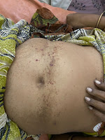
INSPECTION:
1. Shape – distended-uniform
2. Flanks – full
3. Umbilicus – central in Position, Shape-everted (transverse slit)
4. Skin – stretched ,striae present
5. No Dilated veins
PALPATION:
Soft , tenderness diffuse all over the abdomen . No organomegaly
PERCUSSION:
Shifting dullness present
AUSCULTATION:
1. Bowel sounds heard
CARDIOVASCULAR SYSTEM:
S1, S2, heard with no murmurs
EXAMINATION OF RESPIRATORY SYSTEM: NVBS hears.
EXAMINATION OF CNS : NFND
Investigations:
Ascitic fluid amylase - 15
Ascitic fluid protein- 4.1
Sugar- 147
Ascitic fluid LDH- 431
Serology - negative
LFT- TB-0.82,DB-0.20, AST-31,alt- 59,alp-228,
Tp- 5.6, alb-2.2, a/g ratio - 0.66
Fbs-166
Rbs-215
Sr.albumin-2.2
Ascitic albumin-1.7
Saag-0.5
Rft- urea-11,creat-0.7,uric acid - 6.6,ca-8.4,p-5.2, sodium - 138, k-3.9,cl-100
Sr.protein -5.5
Cue-color-reddish
Alb-2+, sugars - nil, pus-10-12,epi cells-3-4, RBC-5-6,others- budding yeast cells present
USG ABDOMEN: gross ascites
Hemogram: hb-9.6, tlc-6700,pcv-28.6, plt-4.38
surgery referral was done in the view of need for peritoneal biopsy.
Advice:plan for diagnostic laparoscopy + proceed to peritoneal or omental biopsy
Report: multiple paracentesis was done with 500 ml draining out each time.
IT WAS A REFRACTORY ASCITES.
17/03/2020:
one unit of PRBC was transfused in view of blood loss during operation and was started on antibiotics post operatively.
INJ.TAXIM /BD
INJ.METROGYL/TID
INJ.AMIKACIN/BD
INJ.PCM/TID
INJ.PAN/OD
Peritoneal biopsy and Omental biopsy:

Microscopy- sections studied from peritoneal biopsy shows chronic inflammatory cell infiltrate in the fibrocollagenous tissue.
sections studied shows lobules of mature adipocytes with area showing chronic inflammatory cell infiltrate comprising of lymphocytes, epithelioid cells and plasma cells.occasional neutrophilic infiltrate seen.Fewmultinucleated gaint cells seen.
Impression- peritoneum- features suggestive of chronic peritonitis.
omentum- features suggestive of granulomatous omentitis.

A drain was placed for 5 days after surgery

20/03/2020:
In the view of above biopsy report patient was initiated on ATT with 3 drugs FDC according to body wt on 20/03/2020 and tab- prednisolone 30mg OD on 20/03/2020.
patient complaints of left hypochondric pain and a review ultrasound and chest x-ray was done.
USG report- Mild to moderate ascitics- echogenic fluid. Rtkidney irregular calycial dilatation noted.
chest x-ray report- Mild to moderate plureral effusion on left side for which
Thoracocentasis was done.

Pleural fluid analysis: sugar-246mg/dl
protein-6.1gm/dl
Pleural fluid analysis : exudative
Ascitic fluid analysis : SAAG : 0.5
Non portal hypertensive gastropathy
DIfferentials. ? TB
? Peritoneal carcinomatosis
? Pancreatitis
GENERAL EXAMINATION
Patient is conscious coherent and cooperative
Built : moderately
Nourishment : moderate
aFebrile
Pallor present
No Icterus Cyanosis Clubbing
Pedal edema and lymphadenopathy
VITAL SIGNS
PULSE: 97 bp regular
BLOOD PRESSURE: 100/70 mm hg supine position right arm
RESPIRATORY RATE : 24 cpm
TEMPERATURE: 98.4 F measured in the Axilla
SYSTEMIC EXAMINATION
ABDOMEN:
INSPECTION:
1. Shape – distended-uniform
2. Flanks – full
3. Umbilicus – central in Position, Shape-everted (transverse slit)
4. Skin – stretched ,striae present
5. No Dilated veins
PALPATION:
Soft , tenderness diffuse all over the abdomen . No organomegaly
PERCUSSION:
Shifting dullness present
AUSCULTATION:
1. Bowel sounds heard
CARDIOVASCULAR SYSTEM:
S1, S2, heard with no murmurs
EXAMINATION OF RESPIRATORY SYSTEM: NVBS hears.
EXAMINATION OF CNS : NFND
Investigations:
Ascitic fluid amylase - 15
Ascitic fluid protein- 4.1
Sugar- 147
Ascitic fluid LDH- 431
Serology - negative
LFT- TB-0.82,DB-0.20, AST-31,alt- 59,alp-228,
Tp- 5.6, alb-2.2, a/g ratio - 0.66
Fbs-166
Rbs-215
Sr.albumin-2.2
Ascitic albumin-1.7
Saag-0.5
Rft- urea-11,creat-0.7,uric acid - 6.6,ca-8.4,p-5.2, sodium - 138, k-3.9,cl-100
Sr.protein -5.5
Cue-color-reddish
Alb-2+, sugars - nil, pus-10-12,epi cells-3-4, RBC-5-6,others- budding yeast cells present
USG ABDOMEN: gross ascites
Hemogram: hb-9.6, tlc-6700,pcv-28.6, plt-4.38
03/04/2020:
E S R # 80 mm/ 1 st hour
HEMOGRAM
HAEMOGLOBIN # 10.0 gm/dl
TOTAL COUNT 5,700 cells/cumm NEUTROPHILS 80 %
LYMPHOCYTES # 15 %
EOSINOPHILS 02 %
MONOCYTES 03 %
BASOPHILS 00 %
PCV # 35.4 vol %
M C V # 72.7 fl
M C H # 20.5 pg
M C H C # 28.2 % RDW-CV # 20.2 %
RDW-SD 48.8 fl
RBC COUNT # 4.87 millions/cumm
PLATELET COUNT 4.60 lakhs/cu.mm SMEAR
RBC Normocytic hypochromic anemia Light Microscopy
WBC With in normal limits Light Microscopy
PLATELETS Adequate in number and distribution Light Microscopy
HEMOPARASITES No hemoparasites seen Light Microscopy
IMPRESSION Normocytic hypochromic anemia
LFT
Total Bilurubin 0.62 mg/dl
Direct Bilurubin # 0.22 mg/dl
SGOT(AST) 20 IU/L
SGPT(ALT) 22 IU/L
ALKALINE
PHOSPHATE
# 232 IU/L
TOTAL PROTEINS # 6.0 gm/dl
ALBUMIN # 2.0 gm/dl
A/G RATIO 0.52
04/04/2020:
C-Reactive Protein -Negative mg/dl
09/04/2020:
HEMOGRAM
HAEMOGLOBIN # 10.5 gm/dl
TOTAL COUNT 5,200 cells/cumm NEUTROPHILS 80 %
LYMPHOCYTES # 16 %
EOSINOPHILS 02 %
MONOCYTES 02 % BASOPHILS 00 % PCV # 35.1 vol %
M C V # 72.9 fl M C H # 21.8 pg
M C H C # 29.9 %
RDW-CV # 20.8 %
RDW-SD 49.3 fl
RBC COUNT # 4.81 millions/cumm
PLATELET COUNT 3.22 lakhs/cu.mm
SMEAR
RBC Microcytic Hypochromic Light Microscopy
WBC With in normal limits Light Microscopy
PLATELETS Adequate in number and distribution Light Microscopy
HEMOPARASITES No hemoparasites seen Light Microscopy
IMPRESSION Microcytic Hypochromic
LFT
Total Bilurubin 0.56 mg/dl Direct Bilurubin # 0.24 mg/dl SGOT(AST) 17 IU/L SGPT(ALT) 13 IU/L ALKALINE
PHOSPHATE
# 203 IU/L TOTAL PROTEINS 7.2 gm/dl ALBUMIN # 2.91 gm/dl
A/G RATIO 0.68
RFT
UREA # 10 mg/dl
CREATININE 0.6 mg/dl
URIC ACID # 8.7 mg/dl DHBS
CALCIUM 9.6 mg/dl
PHOSPHOROUS 3.9 mg/dl
SODIUM 139 mEq/L
POTASSIUM 4.0 mEq/L
CHLORIDE 101 mEq/L
CELL COUNT PLEURAL FLUID
VOIUME 3 ML
COLOUR Pale Yellow
APPEARANCE Turbid
TOTAL COUNT 50 Cells/cumm
DIFFERENTIAL COUNT
NEUTROPHILS 0%
LYMPHOCYTES 100% R B C Nil
OTHERS Nil
LDH
LDH 269 IU/L
PLEURAL (SUGAR, PROTEIN)
SUGAR # 246 mg/dl
PROTIEN 6.1 g/dl .
SERUM PROTEIN
Treatment given :
Tab.pcm 650mg qid
Tab.pan 40mg od
Inj.diclofenac im sos
Due course patient developed arthralgia due to pyrizinamide (ATT) due to hyperuricemia so eventually allopurinol was added and subjectively patient is feelimg better and now presently is arthralgia is it enthesitis due to the same etiology causimg pleuritis serositis is not know.
After 6 months of ATT she was subjectively feeling better and objectively there was no ascites

Comments
Post a Comment