56 year old woman with chest pain since 2020 january presented with pericardial effsuion secondary to multiple myeloma
56 year old female kTobacco leaf seller by occupation came to opd in january 2021 with chief complaints of
chest pain since 6 months , pricking type, subsiding on its own, no radiation
Shortness of breath grade 2(MMRC) since 1 month progressed to grade 4 since 1 day
H/o orthopnoea and Paroxysmal dyspnoea and palpitations present
No history of cough , wheeze
Decreased appetite since 20 days,
generalised weakness since 15 days ,
Pain abdomen since 10 days. Diffuse predominantly in the epigastric region, non radiating
B/L Lower limb ,upper limb edema and facial puffiness since 5 days.
Edema initially started in the lower limbs then prgressed to upper limbs and then face
Palpitations + Syncopal attacks -
She is not a K/C/O DM - II , HTN , Thyroid abnormalities and any other comorbidites
She is an occasional alcoholic ( beer / whiskey) since 35 years. Chews tobacco leaves daily since 6 months.
O/E:
Patient is C/C
Pallor +
No icterus, clubbing , cyanosis, lymphadenopathy
pedal edema present
Temp - 98.6 F
PR - 108 bpm
BP - 120/70 mm of Hg
Spo2 - 98% room
CVS - Inspection :
JVP : Raised JVP prominent X descent - 14 cms.
No precordial bulge or any scars sinuses present
Apical impulse in 6th intercoastal space lateral to mid clavicular line
Palpation
Apical impulse in 6th intercoastal space lateral to mid clavicular line
Parasternal heave +
Ausculation S1 , S2 + muffled heart sounds
Loud P2 in Pulmonary area.
Pansystolic murmur in Tricuspid area.
RS - BAE + , fine inspiratiry crepts present in right and left ISA
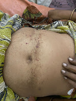
P/A - distended , multiple hemorrhagic spots seen. Hepatomegaly present with liver span of 15 cm
Tenderness + in epigastric and Right hypochondric area.
Provisional diagnosis of Heart failure was made
Blood Investigations

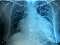
Xray showing money bag appearance indicating pericardial effusion


ESR = 110 mm hg
Suddenly patient developed hypotension,
In view of pericardial effusion she has ,diagnosis of Cardiac tamponade was made
Pulsus pardoxus present
Raised jvp
Muffled heart sounds present
2d echo showed RV diastolic collapse
So immediately pericardiocentasis was done and about 300 ml of fluid was drained
Patient gained a sympotomatic relief but immediately after 20 minutes patient has loss of consciousness
Bp : 60/40 mm hg
Immediately IVF one unit 0.9% NS was given as bolus and inj Noradrenaline at 4 ml/hr was started (1 ml = 80 mcg) according to the dilution
Patient regained consciousness and bp was maintaininv at MAP of 60 mm hg and slowly noradrenaline was tapered.
Pericardial fluid analysis was sent and according to lights criteria , it was exudative
Pericardial fluid for Acid Fast bacilli negative and malignant cells negative
Etiology of pericardial effusion was thought over and considering Tuberculosis as main cause of etiology
Malignancy and Tb were thought over
Thyroid profile was sent which was subclinical hypothyroidism and started on Tab Thyronorm 50 mcg od
Considering Myeloma defining events : anemia , hypercalcemia.
Considering the high Gamma gap serum protein electrophoresis was sent.
Usually the major part of the total protein in out body is imparted by serum albumin . Like wise low serum albumin low protein but this patient of ours has high total protein and low albumin which meant that there are other proteins in the body but not serum albumin Giving us a clue of increased globulins in the body.
Serum protein electrophoresis showing M spike at Beta globulin region

Bone marrow biopsy was done to look for the percentage of plasma cells which were more than 30%

Serum free light chain assay

Treatment
Inj Bortezomib 2mg Sc /od
Inj Cyclophosphamide 400 mg in 500 ml NS Iv /od
These chemotherapeutic agents were given once every 15 -20 days
Maintanince therapyXray after 6 months of treatment

Resolution of pericardial effusion
Patient is on chemotherpay once in 15 days and subjectively she is feeling better
Regained her appetite , no complaints of SOB
Patient is on follow up with Hemogram(Hb rose to 11gm/dl) , LFT recieving Chemotherapy.
Her last chemotherapy was done in the month of october.
She missed her chemotherapy session in november and when they planned for chemosession in december she had pain in the right eye
Going into the details : Patient was apparantly asymptomatic till 25th Decemenber 2021 then she developed pain in the right eye after she applied ointment(?zinda thilasmath) on her right side of forhead and by next morning when she woke up there was redness around the orbit , watering of eyes, pain and tenderness around the right eye.

Patient also complained of fever and slowly patient was in altered sensorium
A working diagnosis of orbital cellulits with Acute kidney injury (Pre -renal /Renal) and uraemic encephalopathy was done and patient underwent 3 sessions of hemodialysis and was started on Antibiotics
Patient sensorium improved and has become subjectively better and was discharged .
From January 8th 2022 patient started developing erythematous lesions on calf region and associated with fever
According to patient attender patient had similar lesions with small distribution whenever there was delay in chemotherapy so these lesions were because she didnt undergo chemotherapy since 2 months.

Discussion
Case 1 A 45-year-old female presented with multiple purpuric, ecchymotic lesions over periorbital area, cheeks, front and back of the neck with relative sparing of covered parts for the last 2 years, with a history of minor trauma precipitating these lesions [Figure [Figure1a1a and andb].b]. Skin was easily fragile in most areas. There was a history of multiple episodes of diarrhea and fever in the past 6 months. General examination showed mild pallor and bilateral pedal edema. Routine investigations are detailed in Table 1. Skin biopsy finding was suggestive of a bullous lesion with amorphous eosinophilic amyloid deposits [Figure 2]. Serum protein electrophoresis (SPE) and bone marrow aspiration (BMA) confirmed it to be a case of MM [Figures [Figures33 and and4].4]. However, radiological evaluations of skeleton were normal. The patient was referred to the department of clinical hematology where chemotherapy was started.
https://www.ncbi.nlm.nih.gov/pmc/articles/PMC5122285/


When was the last follow up?
ReplyDelete